
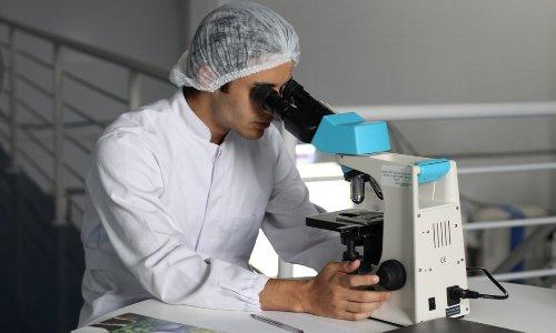
The microscope is seen as an instrument used to inspect objects that are generally too small to be seen with the naked eye. The area of study which involves the investigating of such small objects using a microscope or similar instrument is known as microscopy. The term microscopic is generally used to describe an object invisible to the naked eye unless otherwise assisted by a microscope.
There are several types of microscopes used today. The most common and also first to be invented is known as the optical microscope which uses visible light to image the sample via one or more lenses to generate an enlarged image of a sample positioned in the focal plane and contain a refractive class and often a quartz of plastic yo focus the light towards the eye or another light detector.
Some of the alternative and widely used microscopes include various types of the scanning probe microscope, the ultramicroscope and the electron microscope which includes the scanning electron microscope as well as the transmission electron microscope.
Facts About the Microscope
▪ The optical microscope was the first microscope ever invented, however the inventor to date has never been fully identified. Research nonetheless suggests that the first compound microscope seen in the Netherlands during the late 1500s was likely an invention of the makers of eyeglass there known as Hans Lippershey known for developing the early telescope and Zacharias Janssen who also asserted himself as the inventor of the telescope. Subsequent and unsubstantiated claims were also made along the lines that it was Roger Bacon who invented the telescope and microscope. However it was Giovanni Faber who first conceived the name microscope in 1625 for the compound microscope by Galileo Galilei.
▪ The first comprehensive description of the interior formation of living tissue built on microscopic observation was not seen until 1644, by Italian astronomer Giambattista Odierna however microscopic research was not used extensively for research in Italy, England or the Netherlands until the 1660s and 1670s. Notably it was Italian Marcelo Malpighi who first began the study of biological structures first beginning with the lungs however it was Robert Hooke’s historic book Micrographia detailing the then thirty-year old observations through various lenses and published in September 1655 which had a great impact primarily due to its impressive illustrations.
▪ The most significant contribution to the development of the microscope came from Antonie van Leeuwenhoek on October 9, 1676 who reported the finding of micro-organisms through his discovery of spermatozoa and red blood cells aiding the popularizing of microscopy as an effective approach.
▪ A key technique used for sample illumination and a primary ingredient to modern light microscopy known as Kohler illumination, was developed by August Kohler in 1893. This type of sample illumination was used to later give rise to exceptionally even lighting thus overcoming a vast number of limitations often seen in older techniques of sample illumination. In 9153 new developments in sample illustration was achieved by Frits Zernike’s Phase Contrast and Georges Nomarski development of differential interference contrast illumination in 1955 both of which allowed the imaging of transparent samples which were not stained.
▪ The significant alternative to light microscopy was developed during the early 1900s which saw the use of electrons instead of visible light to produce the images. The first electron microscope, the transmission electron microscope was developed by Ernst Ruska in 1931 and used a similar principle to the optical microscope however used electrons rather than light, and electromagnets instead of glass lenses. The principle was widely accepted as the use of electrons instead of light allowed for much higher resolutions in the images generated. This development of the electron microscope was rapidly succeeded by the evolution of the scanning electron microscope by Max Knoll in 1935.
▪ During World War II electron microscopes became increasingly popular with Ernst Ruska developing the first ever commercial transmission electron microscope while working at the German engineering company known as Siemens ushering a series of scientific conferences on electron microscopy convened during the 1950s.
▪ University of Cambridge Professor of Electrical Engineering Sir Charles William Oatley along with his postgraduate student Gary Stewart developed the first commercial scanning electron microscope labeling the Cambridge Instrument Company as the “Stereoscan”.
▪ The first scanning tunneling microscope was developed by Gerd Binnig and Heinrich Rohrer in 1981. Later followed by the invention of the atomic force microscope by Gerd Binnig, Quate and Gerber.
▪ The most recent progress in light microscopy occurring during the last decades of the 20th century primarily the post-genomic age was primarily centered on the increase of fluorescence microscopy used in biological studies using a a number of techniques one of which includes small chemical staining of cellular formation. This increase in fluorescence microscopy paved the way for one of the major designs in modern microscopy with the development of the confocal microscope. This concept was later patented by Marvin Minsky in 1957 despite the fact that laser technology limited a realistic approach. It was not until the development of the first functional confocal laser scanning microscope by Christoph and Thomas Cremer in 1978 was the technique seen as gaining increased popularity during the 1980s.
▪ Most of the research performed in the early 21st century on techniques aided by the optical microscope is centered on the invention of superresolution (SR) techniques than enhance the resolution of an imaging system for the study of samples labeled via fluorescence.
▪ Microscopes are divided into several varying classes. One class based on whatever interacts with the sample to create the visible image, whether its light of photon used in the optical microscope, electrons used in the electron microscope or a probe which is utilized by the scanning probe microscope etc.
Microscope Webcam
Founded in 2007 Livestream broadcasting to millions of viewers live in HD has served to democratize live video broadcasting by providing individuals with the right tools to stream almost any event online.
The website features a live webcam showing microscopic life forms as seen in a drop of water shown in the link posted below.
View Under-microscope.com web cam.